DISCITIS
Delving Into the Complexities of Several Spinal Inflammations
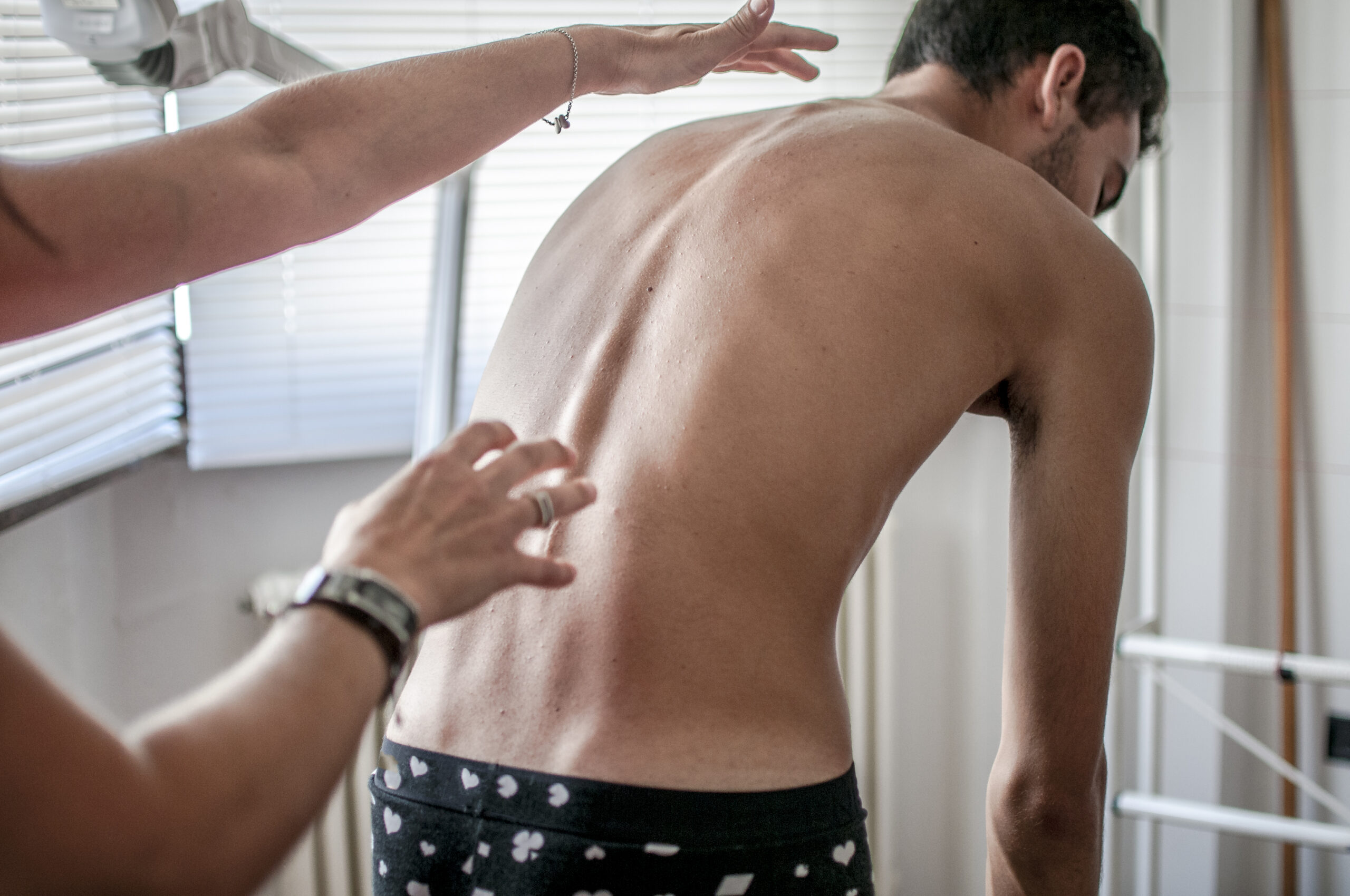
Discitis, or Diskitis, is one of several forms of spinal inflammation characterized by inflammation of the intervertebral disc spaces, which serve as cushiony pads between the vertebrae in the spinal column. This condition can lead to severe back pain and discomfort (1).
While discitis is relatively uncommon, it can affect adults as well, although it is predominantly observed in children. Furthermore, it is often diagnosed alongside another condition known as osteomyelitis, which involves inflammation of the vertebral bone itself (2).
Diskitis affects somewhere between 1 in 100,000 and 1 in 250,000 people in the United States. Interestingly, the prevalence of diskitis is similar in other developed countries, but infectious diskitis is notably more common in less developed nations. Notably, as much as 11% of back pain patients in some African regions receive a diskitis diagnosis (3, 4).
In adults, males exhibit a higher prevalence of diskitis, with male-to-female ratios ranging from 2:1 to 5:1. However, childhood diskitis shows a slight male preponderance, with a male-to-female ratio of 1.4:1 (5).
Symptoms of Discitis
The most common symptom of Discitis is back pain in a specific area of the spine. The intensity of the pain maybe mild or severe, but can become worse when moving or when pressure is put on the affected area. In addition, other symptoms of Discitis include:
-
- Stiffness of the back
- Posture change
- Pain or discomfort in the abdomen
- Difficulty in performing regular mobility tasks
- Loss of appetite
- Radiating pain
- Fever
- Malaise (3)
It’s important to be aware of these potential symptoms and consult with a healthcare professional for proper evaluation and diagnosis if any of these signs persist
What causes Discitis?
Infection, frequently initiated by bacteria and occasionally by fungi, is the primary culprit behind discitis. Specifically, Staphylococcus aureus is the bacterium most commonly pinpointed, while Escherichia coli, Streptococcus pneumoniae, and Salmonella spp. are among the other formidable microorganisms that can lead to discitis (6).
In addition to infections, discitis can also be incited by autoimmune conditions such as rheumatoid arthritis, where the body’s immune system erroneously attacks and damages its own tissues (7). Furthermore, accidents or spinal surgery can lead to spontaneous discitis, typically manifesting as a post-operative complication (8). Although less common, discitis can also arise due to age-related deterioration of the intervertebral discs (9).
Diagnosis
Physicians usually diagnose Discitis using a combination of clinical evaluation, imaging studies and laboratory tests. The imaging studies which may be conducted are:
- MRI (10)
- CT scans (11)
- X-rays (9)
Besides these, the laboratory tests routinely undertaken to detect Discitis are:
- Blood Tests
Inflammatory indicators such as C-reactive protein (CRP) and erythrocyte sedimentation rate (ESR) may be high in patients with Discitis, indicating an inflammatory process (2).
- Microbial Cultures
If bacterial or fungal infection is suspected, blood cultures or biopsy samples from the afflicted disc may be taken in order to identify any infectious agents (11).
How do Physicians Treat Discitis?
Treatment for Discitis is individualized and tailored to each person, taking into account the underlying cause of the condition, any coexisting health issues, and the severity of symptoms.
Medical Therapy
- Physicians typically prescribe specific antibiotics to treat bacterial discitis, with the choice of antibiotics guided by the results of microbial culture and sensitivity tests. The duration of the antibiotic course can vary but often spans 4-6 weeks.
- In cases where the infection is fungal in nature, healthcare providers may recommend anti-fungal medications like Fluconazole.
- Additionally, analgesics or pain medications may be prescribed alongside antibiotic therapy to alleviate discomfort, improve mobility, and facilitate daily activities (6, 12).
Bracing and Immobilization
In the early stages of the disease, it’s important to maintain stillness to facilitate the proper healing of the affected spinal bones. Therefore, physicians often recommend that patients begin with two weeks of bed rest. Subsequently, they should transition to wearing a brace while gradually resuming movement. Braces are typically worn for a duration of 3 to 6 months after initiating treatment (2).
Surgical Management
Generally, early detection can obviate the need for surgical intervention. However, in severe cases, surgery may become necessary, particularly for patients with neurological impairment or spinal instability (12). Two viable surgical procedures include surgical debridement of the infected disc and spinal fusion for stabilization (13).
The Key to Thriving
- Pain Management: Effective pain management is crucial for chordoma recovery. This involves ongoing medication management, making lifestyle adjustments, and exploring alternative pain management techniques.
- Proactive Monitoring: It’s essential to be proactive about one’s health by following prescribed treatments, managing coexisting medical conditions, and promptly reporting any new or worsening symptoms to the healthcare team.
Believe you have symptoms of Discitis?
Contact Longhorn Brain & Spine
Bibliography
- Muscara JD, Blazar E. Diskitis. StatPearls [Internet]: StatPearls Publishing; 2023.
- Gouliouris T, Aliyu SH, Brown NM. Spondylodiscitis: update on diagnosis and management. Journal of Antimicrobial Chemotherapy. 2010;65(suppl_3):iii11-iii24.
- Cottle L, Riordan T. Infectious spondylodiscitis. Journal of Infection. 2008;56(6):401-12.
- Bileckot R, Ntsiba H, Okongo D, Ognami J. Diagnosis of arthritis in black Africa. Apropos of 473 cases in Congo. Revue du Rhumatisme (Ed Francaise: 1993). 1994;61(4):260-5.
- Lam KS, Webb JK. Discitis. Hospital Medicine. 2004;65(5):280-6.
- Principi N, Esposito S. Infectious discitis and spondylodiscitis in children. International journal of molecular sciences. 2016;17(4):539.


