SPINAL TUMORS
What Is a Spinal Tumor?
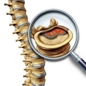
The medical term “Spinal Tumor” refers to the abnormal cell growth that develops within or surrounding the spinal cord or spinal canal. These cells usually grow and multiply uncontrollably, with the two most common classifications including benign (non-cancerous) or malignant (cancerous). Besides these, primary tumors develop within the spine or spinal cord and secondary or metastatic tumors originate outside of the spinal cord and then spread to the spine. The bones, nerves, and other tissues that make up the spine can develop tumors. Only around 10% of spine tumors originate in the spine (Kumar, Tan, Wei, & Vellayappan, 2020). Low grade spinal malignant tumors grow slowly and high-grade spinal tumors grow rapidly.
The classification of the spinal tumor depends on the location of tumors within the spine. The 3 main types of spinal tumors include intradural-extramedullary, intramedullary, and extradural. In addition, these tumors can occur in the cervical, thoracic, lumbar, and sacrum regions of the spinal cord. Intradural-extramedullary spinal tumors grow inside of the covering of the spinal cord (the dura), but outside the actual spinal cord. This site comprises almost 40% of the frequency of occurrence. The most frequent of these cancers form in the arachnoid membrane of the spinal cord (meningiomas), in the nerve roots that branch out from the spinal cord (schwannomas and neurofibromas), or at the spinal cord base (schwannomas and neurofibromas). Meningiomas can reoccur and neurosurgeons find them very difficult to remove. Ependymomas at the end of the spinal cord can be big, and removal can be difficult due to the delicate nature of small neuronal structures in that area (Gerszten & Welch, 2004).
Intramedullary tumors grow inside the spinal cord and usually derive from glial or ependymal cells found throughout the interstitium of the spinal cord. The most common types of intramedullary tumors include astrocytoma and ependymomas, which comprise a 5% occurrence rate. Lastly, extradural tumors originate outside of the dura, which covers the spinal cord as a thin membrane. Common pediatric spinal tumors may include osteoid osteoma, osteosarcoma, osteoblastoma, chordoma, fibroma, hemangioma, mesenchymal Chondrosarcoma, angiosarcoma, etc.
What Are the Common Signs and Symptoms of a Spinal Tumor?
The signs and symptoms of spinal tumors depend upon the growth rate and location of the tumors. The growth of a spine tumor can weaken bones and pressure the spinal cord and nerves, resulting in spinal fractures and neurologic damage. The first signs of developing a spinal tumor include discomfort and severe pain in a patient. The type of discomfort can reveal a lot about the origin of the tumor.
Pain that occurs primarily during sitting or standing indicates that the tumor causes weakness of the spine’s bones and results in spinal instability. The pain usually starts in the early morning or night and patients usually feel better as they move. This pain occurs because of the tumor-induced inflammation while sleeping. Closely located spine tumors can interfere with the ability of major nerves to convey messages between the body and the brain. This can result in neurologic symptoms, such as (Ciftdemir, Kaya, Selcuk, & Yalniz, 2016)
- Walking difficulties
- Problems with balance and coordination
- Problems with sensory functions
- Loss of sensitivity to pain, heat, and cold
- Loss of bladder and bowel control
- Tingling, numbness, and weakness in the limbs
- Pain at the site of the tumor
How Do Spinal Specialists Diagnose Spinal Tumors?
Not all back pain indicates a spinal tumor. For accurate diagnosis of spinal tumors, patients should undergo a thorough medical examination performed by a neurosurgeon. This examination should emphasize the back pain and neurological deficits of the patient. Neurosurgeons further perform radiology tests to confirm their diagnosis. These includes (Reith, 2011):
- Computed tomography (CT) scan: This test helps to visualize the shape and size of the spinal canal, its contents, and the surrounding structures.
- X-ray: This also helps to visualize the structure of the vertebrae and the outline of the joints and helps the neurosurgeon to see any abnormalities within or surround the spinal cord.
- MRI: This produces three-dimensional images of body structures and helps to detect any abnormalities in the spinal cord.
- Bone scan: This test uses Technetium-99 to identify any bony tumors, bone infection, and diseases involving abnormal bone metabolism.
- Biopsy: This includes a neuropathologist cutting the tumor into small pieces to diagnose the type.
How Do Spinal Specialists Treat Spinal Tumors?
Treatment procedure of a spinal tumor depends upon the type, size, and location of the spinal tumor (Li et al., 2021). Neurosurgeons may perform non-surgical options such as radiation therapy, chemotherapy, etc., or surgical options to treat spinal tumors. At the initial stage, some tumors respond well to the non-surgical options. Most metastasis tumors become radioresistant and require surgical options to remove the cancerous cells. Spinal Specialists may suggest surgery if patients display intractable pain, spinal cord compression, and spinal fractures. The risks of spinal tumor surgery can include excessive bleeding, risk of infection, hematoma, etc. For more information, please contact us with any questions you may have..
References
Ciftdemir, M., Kaya, M., Selcuk, E., & Yalniz, E. (2016). Tumors of the spine. World Journal of Orthopedics, 7(2), 109–116. https://doi.org/10.5312/wjo.v7.i2.109
Gerszten, P. C., & Welch, W. C. (2004). Cyberknife radiosurgery for metastatic spine tumors. Neurosurgery Clinics of North America, 15(4), 491–501. https://doi.org/10.1016/j.nec.2004.04.013
Kumar, N., Tan, W. L. B., Wei, W., & Vellayappan, B. A. (2020). An overview of the tumors affecting the spine – Inside to out. Neuro-Oncology Practice, 7, I10–I17. https://doi.org/10.1093/nop/npaa049
Li, J., Wei, W., Xu, F., Wang, Y., Liu, Y., & Fu, C. (2021). Clinical Therapy of Metastatic Spinal Tumors. Frontiers in Surgery, 8(April), 1–12. https://doi.org/10.3389/fsurg.2021.626873
Reith, W. (2011). [Spinal tumours]. Der Radiologe, 51(12), 1016–1017. https://doi.org/10.1007/s00117-011-2150-x
ABOUT LONGHORN BRAIN & SPINE
Founded on Excellence
Founded by Neurosurgeon, Dr. Grant Booher, Longhorn Brain and Spine focuses on a patient-centered approach to alleviating North Texans from Neurological and Spinal Pain. Dr. Booher and his clinical team believe in exhausting all non-invasive protocols first and if needed, employing the least invasive procedures necessary to treat the patients.
Our Beliefs
Dr. Booher believes in a conservative, individualized and holistic approach when it comes to his patients. He prefers exhausting all nonsurgical options and proudly offers the least invasive techniques when clinically indicated. He strives to treat every patient like a member of his family. During his free time, he and his wife enjoy watching sports, listening to Texas country music, and traveling.