DTRAX CERVICAL FACET FUSION
What’s Dtrax Cervical Facet Fusion?
Dtrax Cervical Facet Fusion refers to a minimally invasive technique used to carry out posterior fusion of the cervical vertebrae. This procedure involves the use of a set of single-use surgical instruments to drill into, insert a “cage” and then fuse the lateral aspects of two cervical vertebrae. Spine Specialists often perform Dtrax cervical facet fusion in conjunction with anterior cervical decompression and fusion to achieve cervical spinal decompression and stabilization (Krer et al., 2020).
In recent times, minimally invasive techniques have emerged as the preferred means of performing spinal fusion particularly in high-risk older patients with multiple comorbidities previously considered ineligible for traditional open surgical intervention. Dtrax Cervical Facet Fusion has demonstrated excellent outcomes in reducing the risk of perioperative and postoperative complications. This innovative, tissue sparing, posterior cervical approach allows for faster recovery time, shorter hospital stay, and negligible blood loss (Laratta, Gupta & Smith, 2020).
In addition, research has proven that minimally invasive posterior cervical fusion with facet cages yields similar stability to traditional open posterior cervical fusion surgery with lateral mass screw (Kramer et al., 2020).
When is Dtrax Cervical Facet Fusion Recommended?
The indications of Dtrax cervical facet fusion include cervical radiculopathy from;
However, before neurosurgeons carry out the procedure, the patient has to meet certain criteria (McCormack et al., 2013). Some of the criteria include;
- The affected cervical vertebra must occur within the C3-C5 levels.
- Only skeletally mature adults can undergo this procedure.
- The patient must have had at least 6 weeks of non-operative conservative management which failed to resolve the symptoms.
When’s Dtrax Cervical Facet Fusion Not Recommended?
The presence of certain clinical conditions makes Dtrax cervical facet fusion difficult or risky to safely and efficiently carry out. Before a decision to conduct this procedure, the neurosurgeon excludes the following (Clinicaltrials.gov, 2020);
- Any active infective process or patients with a significant risk of infection (immunocompromised).
- Pregnancy.
- Local inflammation or infection around the operation site.
- Fever or leukocytosis.
- Morbid obesity.
- Distorted anatomy from congenital anomalies.
- Osteopenia.
- Osteoporosis.
- Cancer of the spine.
What’s the Benefits of Dtrax Cervical Facet Fusion?
Dtrax cervical facet fusion has an excellent tissue-sparing feature. All the instruments used in this procedure have a diameter of less than 1cm. This greatly minimizes damage to muscles and soft tissue along the surgical path. Dtrax cervical facet fusion consequently, leads to –
- Reduced surgery time
- Reduced blood loss
- Reduced hospital stay
- Reduced recovery time
- Reduced postoperative pain
- Reduced scarring
How do patients Prepare for Dtrax Cervical Facet Fusion?
In preparation for the procedure, neurosurgeons often recommend an overnight fast of 8-12 hours. This reduces the risk of anesthetic complications.
Patients need to avoid taking non-steroidal anti-inflammatory medicines (ibuprofen, diclofenac, etc.) and blood thinners (Coumadin, aspirin, Plavix, etc.) for at least a week prior to the surgery.
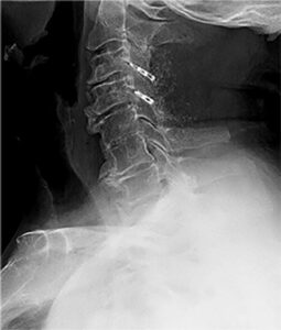
The neurosurgeon would also have the patient undergo imaging studies such as X-rays, CT and MRI scans prior to surgery.
How do Neurosurgeons perform Dtrax Cervical Facet Fusion?
The anesthesiologist will put the patient under anesthesia and monitor their vital signs during the surgery.
The patient will lie prone on the operating table. The neurosurgeon will perform this procedure on the back of the patient’s neck hence the need to lie face down. Afterwards, an assistant sets up the appropriate imaging machine to aid visualization.
The neurosurgeon identifies the exact level affected with the aid of a spinal needle using fluoroscopy and draws corresponding surface markings on the skin of the patient to indicate landmarks. The neurosurgeon will then place a surgical incision about 4cm below the affected level.
Following this, the surgeon uses the Dtrax single-use surgical instruments (all smaller than one centimeter in diameter) to prepare the bony surfaces of the cervical spine for fusion.

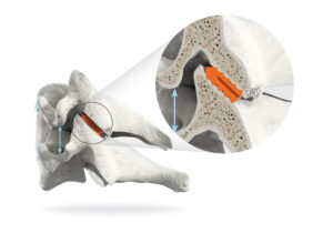
The neurosurgeon will insert an appropriate sized Dtrax cervical cage prefilled with bone graft into the space between the superior and inferior cervical facets and screw the cage in place using the Dtrax cervical screw. This creates more space for a pinched or irritated nerve root while also stabilizing the spine allowing the fusion to heal properly (Laratta, Gupta & Smith, 2020). Finally the surgeon adds more bone graft around the cage and withdraws the instruments and sutures the skin.
The neurosurgeon performs this procedure bilaterally on either side of the cervical facets to achieve a balanced fusion.
What does Recovery after Dtrax Cervical Facet Fusion look like?
In most cases, the neurosurgeon discharges the patient on the same day. Recovery varies from person to person but most patients return to their regular activities within a week.
What’s the Possible Complications of Dtrax Cervical Facet Fusion?
Rarely any complications occur with cervical facet fusion using Dtrax. However, possible complications may include; surgical site infection and fracture of the facet during insertion of the implant.
References
- Kramer, S., Albana, M. F., Ferraro, J. B., & Shah, R. V. (2020). Minimally Invasive Posterior Cervical Fusion with Facet Cages to Augment High-Risk Anterior Cervical Arthrodesis: A Case Series. Global spine journal, 10(2 Suppl), 56S–60S. https://doi.org/10.1177/2192568220911031
- Laratta, J. L., Gupta, K., & Smith, W. D. (2020). Tissue-Sparing Posterior Cervical Fusion with Interfacet Cages: A Systematic Review of the Literature. Global spine journal, 10(2), 230–236. https://doi.org/10.1177/2192568219837145
- McCormack, B. M., Bundoc, R. C., Ver, M. R., Ignacio, J. M., Berven, S. H., & Eyster, E. F. (2013). Percutaneous posterior cervical fusion with the DTRAX Facet System for single-level radiculopathy: results in 60 patients. Journal of neurosurgery. Spine, 18(3), 245–254. https://doi.org/10.3171/2012.12.SPINE12477
- Clinicaltrials.gov (2020), Evaluation of Cervical Fusion with DTRAX® Cervical Cage with DTRAX Bone Screw, NCT02694250. https://clinicaltrials.gov/ct2/show/study/NCT02694250