FLOROSCOPY
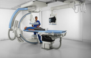
Fluoroscopy refers to an imaging technique that uses X-rays to obtain real-time moving images of the interior of an object. Fluoroscopy includes two major sub-categories. Larger, typically floor, wall or ceiling mounted device often called Cath Lab, and smaller (but categorized as full size and mini C-Arm) Mobile C-Arm. In its primary application of medical imaging, a fluoroscope allows a surgeon to see the internal structure and function of a patient mainly during surgery so that the surgeon can watch the pumping action of the heart, or the motion of swallowing, for example.
This proves useful for both diagnosis and therapy and occurs in general radiology, interventional radiology, and image-guided surgery.
Surgeons typically use mobile C-Arm (Full Size & Mini C-Arm) for surgical and pain management in many different fields including vascular, orthopedic, podiatry, urology, fertility clinic, and many other areas that require use of live X-ray. While some Mobile C-Arms prove useful for vascular cases, most cases for Mobile C-Arm include the ability to see structures (spine and other bones) for injection or minimally invasive surgical procedures. Full Size and Mini C-Arm mainly differ based on what object/interest these devices target. For extremity procedures (hands and foot), a Mini C-Arm can do the job while shoulder, hips and spine may require bigger sized, Full Size C-Arm.
In its simplest form, a fluoroscope consists of an X-ray source and a fluorescent screen, between which the surgeon places the patient. However, since the 1950s, most fluoroscopes have included X-ray image intensifiers and cameras as well, to improve the image’s ability and make it available on a remote display screen. For many decades, fluoroscopy tended to produce live pictures that did not record, but since the 1960s, as technology improved, recording and playback turned into the norm.
Fluoroscopy shares certain similarities with radiography and X-ray computed tomography (X-ray CT) in that it generates images using X-rays. The original difference included the fact that radiography fixed still images on film, whereas fluoroscopy provided live moving pictures that did not store. In recent times, radiography, CT, and fluoroscopy all refer to digital imaging modes with image analysis software and data storage and retrieval.
Medical Applications
Fluoroscopy has proved an important tool in medical imaging to render moving pictures during a surgery or any other procedure.
- Cardiology
In cardiology, specialists use fluoroscopy for diagnostic angiography, percutaneous coronary interventions, implantable cardioverter defibrillators, pacemakers, and cardiac resynchronization devices.
- Surgical fluoroscopy
Surgeons use fluoroscopy in various types of surgical procedures, such as orthopedic surgery and podiatric surgery. In both of these procedures, fluoroscopy aids in reducing the risk of fractures and in certain procedures that have extensive hardware.
- Urology
In urology, specialists use fluoroscopy in retrograde pyelography and micturating cystourethrography to detect various abnormalities related to the urinary system.
- Gastrointestinal fluoroscopy
In gastrointestinal examinations, specialists use fluoroscopy to examine the digestive system by using a substance opaque to X-rays (usually barium sulfate or gastrografin). Specialists introduce these substances into the patient’s digestive system either by swallowing or as an enema. This usually acts as part of a double-contrast technique, using positive and negative contrast. Barium sulfate coats the walls of the digestive tract (positive contrast), which allows the specialist to view the outline of the GIT as white or clear on an X-ray. Presence of air (negative contrast) in the digestive tract appears black on the film. The barium meal includes an example of a contrast agent swallowed to examine the upper digestive tract. While soluble barium compounds prove toxic, the insoluble barium sulfate proves nontoxic due to its low solubility that prevents the body from absorbing it. Investigations of the gastrointestinal tract include barium enemas, defecating proctograms, barium meals and swallows, and enteroclysis.
Other medical uses include:
- Angiography of the leg, heart, and cerebral vessels.
- Placement of a peripherally inserted central catheter.
- Discography, an invasive diagnostic procedure for evaluation for intervertebral disc pathology.
- Placment of a weighted feeding tube (Dobhoff for example) into the duodenum after previous attempts without fluoroscopy have failed.
- Specialists perform liver biopsy under fluoroscopic guidance at many centers.
- Lumbar puncture. Fluoroscopy helps to guide where the needles of the spinal tap can go, and may reduce the number of attempts required for a successful lumbar puncture.